If you’ve been referred for a scan, you might be wondering what a gallstones ultrasound actually shows. In simple terms, a gallstones ultrasound can detect solid particles within the gallbladder by identifying how they reflect sound waves. These stones often appear as bright echoes on the scan and may cast shadows behind them. Understanding how an ultrasound to detect gallstones works can help explain why it’s the most common test used to assess gallbladder concerns. Many people are referred for this scan after experiencing symptoms such as abdominal discomfort, particularly after eating.
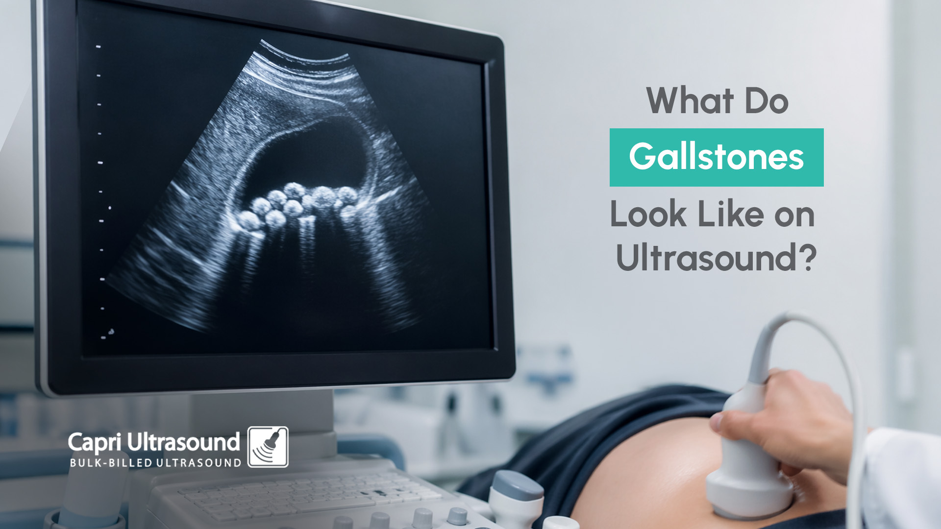
A common question we get a lot is: “what do gallstones look like on ultrasound?”
On a scan, gallstones typically appear as:
This shadowing is one of the key features that helps distinguish gallstones from other findings.
Because ultrasound provides real-time imaging, the sonographer can observe how these structures behave, which helps confirm whether they are stones.
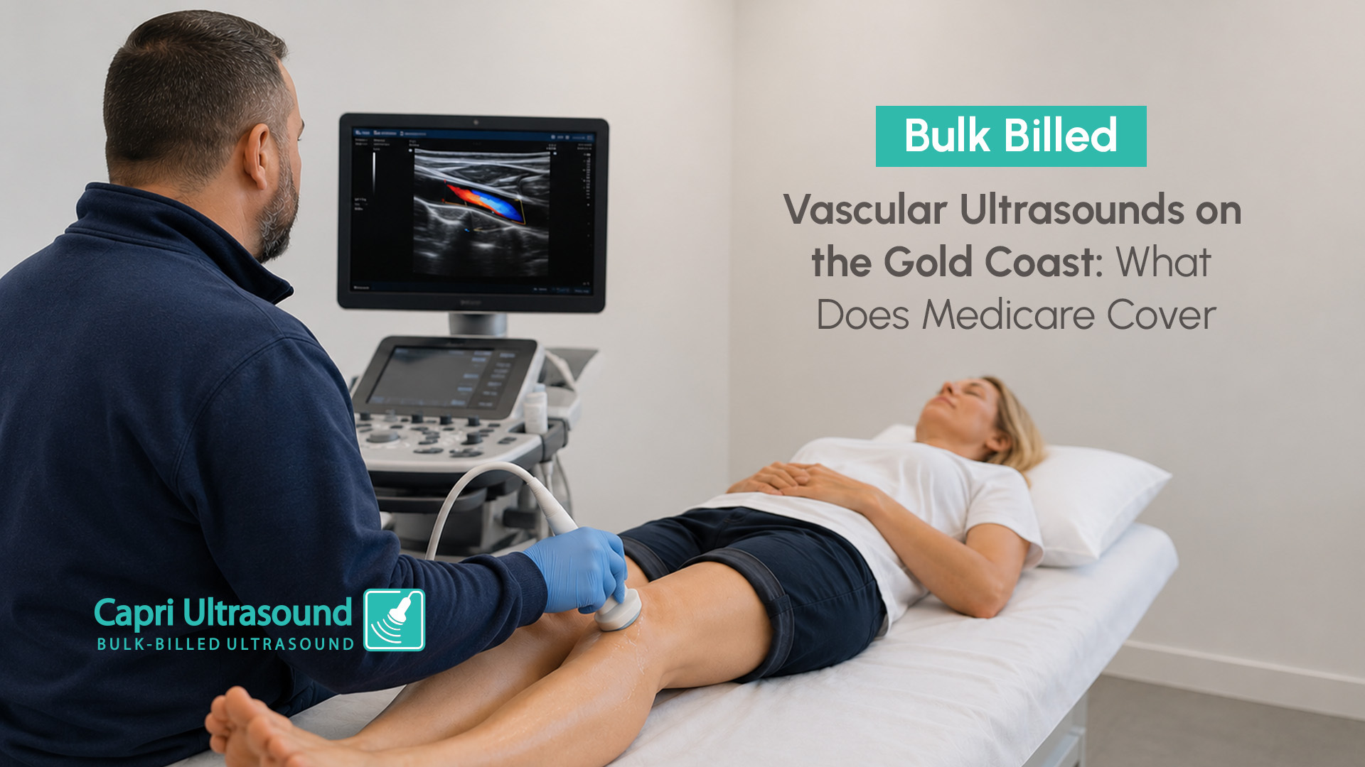
When performing an ultrasound of the gallbladder with gallstones, the ultrasound uses high-frequency sound waves to create images of the gallbladder and surrounding structures.
The scan is used to:
This type of imaging is non-invasive and does not involve radiation, making it a common first step when investigating upper abdominal symptoms.
The sonographer is looking for specific features that indicate the presence of stones.
These may include:
In some cases, multiple stones may be present, while in others there may only be a single stone.
Google searches like “ultrasound pictures of gallstones” or “gallstones in ultrasound images” are common, but it’s important to understand that interpretation involves more than just appearance. Movement and shadowing are key factors that help confirm the diagnosis.
To understand abnormalities, it helps to know what a normal gallbladder ultrasound looks like.
In a healthy scan, the gallbladder typically appears:
Comparing this to an ultrasound of gallbladder with gallstones makes it easier to identify when something is not typical.
Another finding that may be seen is gallbladder sludge ultrasound appearances. ‘Sludge’ refers to thickened bile that can appear as low-level echoes within the gallbladder. Unlike stones, sludge does not usually cast a shadow and may shift slowly with movement. While it is different from gallstones, it can sometimes be associated with similar symptoms.
A gallstones ultrasound may be recommended when patients experience symptoms such as:
It is often the first imaging test used because it provides a clear view of the gallbladder and can quickly identify whether gallstones are present.
While many people with gallstones may not have symptoms, in some cases they can lead to complications.
One example is inflammation of the gallbladder, known as cholecystitis, which may cause more persistent pain or discomfort. For a more detailed explanation of this condition, Mayo Clinic provides further information here: Cholecystitis
If your GP has recommended a gallbladder ultrasound to detect gallstones, having the scan performed can help clarify the cause of your symptoms.
Capri Ultrasound, located on the Isle of Capri on the Gold Coast, provides diagnostic imaging including gallbladder ultrasounds to assess gallstones and related conditions.
With a valid referral, under medicare patients are eligible for many bulk billed ultrasounds, making it easier to access appropriate imaging.
If you’ve been experiencing symptoms or have been referred for a gallbladder ultrasound, booking your scan can help provide answers and guide the next steps in your care.
To learn more or make a booking, contact Capri Ultrasound on the Gold Coast today.