
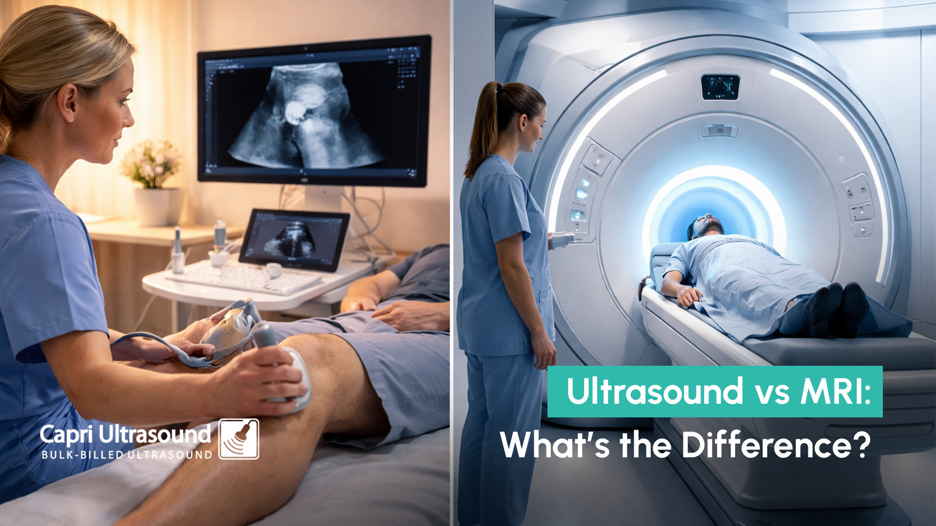
If your doctor has recommended imaging, you might be wondering about ultrasound vs MRI and which one you actually need. In simple terms, both scans produce images of the body, but they work in very different ways and are used for different reasons. Understanding ultrasound vs MRI can help you feel more confident about why a particular scan has been recommended.
In many cases, ultrasound is used as a first-line imaging tool, especially for soft tissue, vascular, and abdominal concerns.
The difference between MRI and ultrasound comes down to how the images are created and what they are best at detecting.
An ultrasound uses high-frequency sound waves to produce real-time images of structures inside the body. It is commonly used to assess muscles, tendons, organs, and blood flow.
MRI (magnetic resonance imaging), on the other hand, uses strong magnetic fields and radio waves to create highly detailed images of internal structures. It is often used for more complex areas such as the brain, spine, and joints.
When comparing ultrasound vs MRI, ultrasound is typically:
MRI is typically:
A common question patients ask is what can ultrasound detect. Ultrasound is a versatile imaging tool used across many areas of medicine.
It can help assess:
In fact, many diseases can be detected by ultrasound, particularly those affecting soft tissues and circulation. Because it provides real-time imaging, it is especially useful for identifying inflammation, fluid collections, and changes in blood flow.
This is why ultrasound is often the first imaging test requested by GPs before considering more advanced scans.
Understanding ultrasound vs MRI also means knowing when ultrasound is the preferred option.
Doctors may recommend ultrasound when:
Ultrasound is often chosen because it can provide immediate information without the need for more complex imaging. For many conditions, it is an effective first step in diagnosis and may reduce the need for further testing.
MRI is generally used when more detailed imaging is required or when ultrasound cannot adequately assess a specific area.
For example, MRI may be recommended for:
MRI scans use powerful magnets to create detailed cross-sectional images. If you’d like to understand more about how MRI works and what it shows, Cleveland Clinic provides a detailed explanation on MRI (Magnetic Resonance Imaging). This type of imaging is often used after an initial assessment or when further detail is needed.
When deciding between ultrasound vs MRI, the choice depends on:
In many cases, ultrasound is used first because it is efficient, accessible, and provides valuable diagnostic information. If additional detail is required, an MRI may then be recommended.
This step-by-step approach helps ensure patients receive the most appropriate imaging without unnecessary tests.
If your doctor has recommended imaging and you’re unsure about ultrasound vs MRI, starting with the right scan can make the process simpler.
Capri Ultrasound, located on the Isle of Capri on the Gold Coast, provides diagnostic imaging including musculoskeletal, abdominal, and vascular scans. This includes doppler ultrasound to assess blood flow and investigate circulation concerns.
With a referral from a doctor, patients are eligible for a bulk billed ultrasound, making it a practical first step in diagnosis.
If you’ve been referred for imaging or are experiencing ongoing symptoms, booking an ultrasound can help provide clarity and guide the next steps in your care.
To learn more or make a booking, contact Capri Ultrasound Gold Coast today.