
Knee pain and injury are common issues among Australians, from athletes and tradies to retirees and office workers. Whether it’s a sudden injury or ongoing discomfort, a knee ultrasound is often one of the first diagnostic tools used to help identify the underlying problem. Safe, non-invasive and relatively quick, this imaging method can provide detailed insights into soft tissue damage, inflammation and fluid build-up all without radiation exposure.
If you’re experiencing persistent knee issues, this blog will help you understand what a knee ultrasound is, what it can detect, when you might need one, and what to expect during the process.
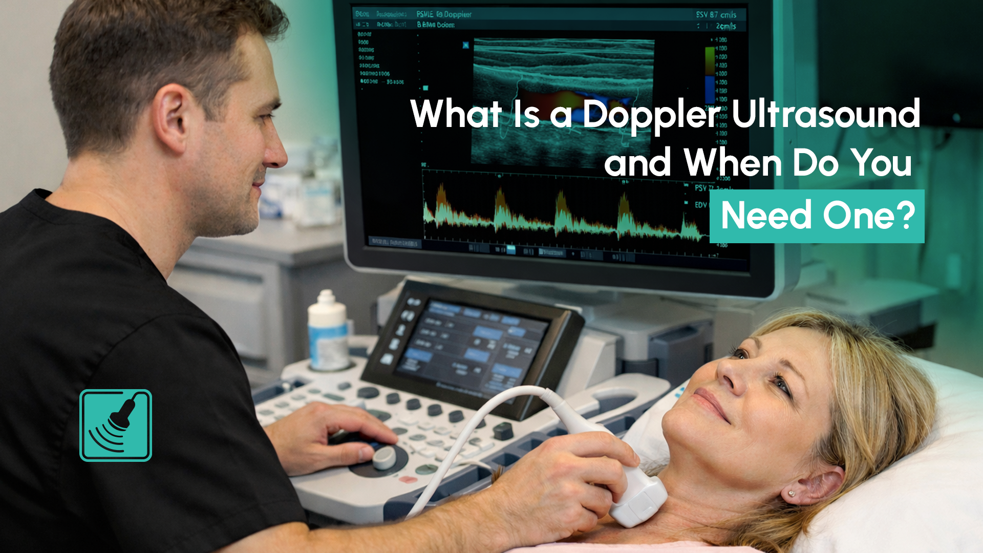
A knee ultrasound (also known as a knee joint ultrasound) is a medical imaging procedure that uses high-frequency sound waves to create real-time images of the soft tissues in and around the knee. Unlike X-rays, which are ideal for viewing bones, ultrasound is best for assessing structures like ligaments, tendons, bursae and fluid-filled areas.
Because the scan is dynamic, the technician may ask you to move or flex your knee during the process, allowing them to assess how the tissues function in motion, something other static scans, like MRI or CT, can’t easily offer.
A knee ultrasound is particularly effective for detecting:
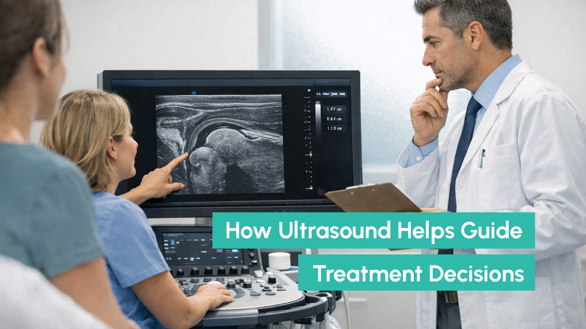
In some cases, your GP may also use the ultrasound findings to guide treatment plans, including cortisone injections or physiotherapy referrals.
If you’re experiencing any of the following, your GP may refer you for a knee ultrasound:
In many cases, early imaging can help prevent further damage and ensure you’re on the right treatment path from the start.
The procedure is straightforward and usually takes between 10 to 20 minutes. Here’s what typically happens:
The scan is painless, and the gel wipes off easily afterwards. There are no side effects or downtime, so you can resume normal activity straight after the appointment.
While a knee ultrasound is excellent for soft tissue assessment and dynamic scanning, it does have limitations. For example, evidence of cartilage damage and meniscus tears is not usually visible on ultrasound and analysis of the cruciate ligaments is not possible.
In such cases, your GP or specialist might recommend an MRI for a more detailed view. MRI is particularly useful for detecting bone bruising, internal ligament ruptures, or early osteoarthritis.
One of the major benefits of early imaging is timely treatment planning. For instance, identifying patellar tendonitis early allows for modification of activity and structured rehab, preventing long-term strain on the joint.
For some patients, even understanding that the pain is due to a mild inflammation (rather than a tear) offers reassurance and avoids unnecessary treatment.
A knee ultrasound is an efficient, safe and highly effective way to diagnose many common causes of knee pain, from sports injuries to overuse conditions and arthritis-related inflammation. If you’ve been struggling with discomfort, clicking, swelling or limited movement, ask your GP whether this type of imaging is right for you.
At Capri Ultrasound, we accept all referrals, and offer bulk billed knee ultrasounds with fast turnaround times and expert diagnostic reporting. Contact us today to book your scan or speak to your GP for a referral to us today!