If your doctor has referred you for an arterial ultrasound, you may be wondering what it actually involves and why it’s necessary. This quick, non-invasive test can provide vital information about the health of your arteries and how well your blood is circulating. It’s commonly used to check for narrowing, blockages, and aneurysms, often before symptoms even appear. Whether you’re experiencing high blood pressure, dizziness, or have a family history of vascular issues, an arterial ultrasound can help uncover problems early and guide your next steps.
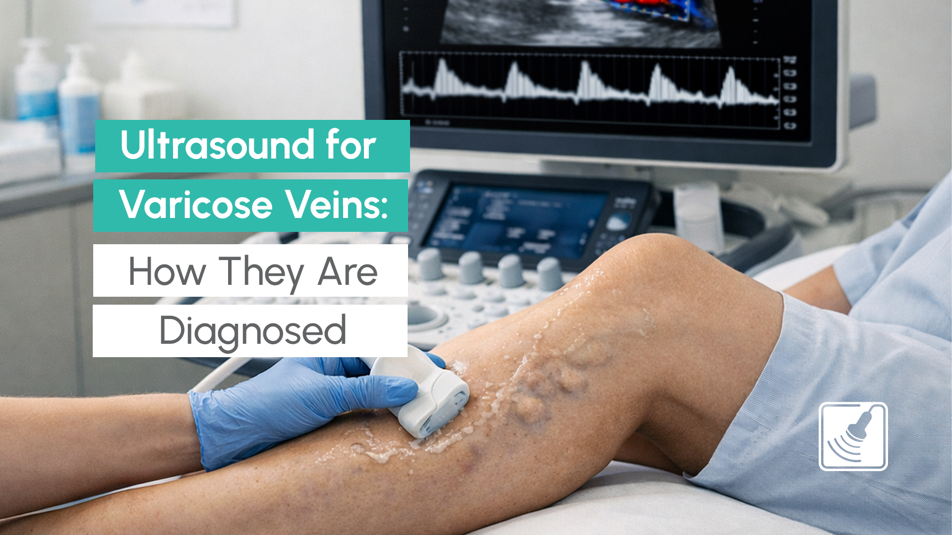
An arterial ultrasound is a diagnostic imaging scan that uses high-frequency sound waves (ultrasound) to assess blood flow through your arteries. It’s often performed with Doppler ultrasound technology, which allows the sonographer to measure the speed and direction of blood as it flows through your vessels.
The test is painless and doesn’t involve radiation, needles, or contrast dyes. It can be done on various arteries in the body, including those in the neck (carotid arteries), abdomen, legs, and kidneys, depending on what your doctor is investigating.
Carotid Artery Disease & Stroke Risk
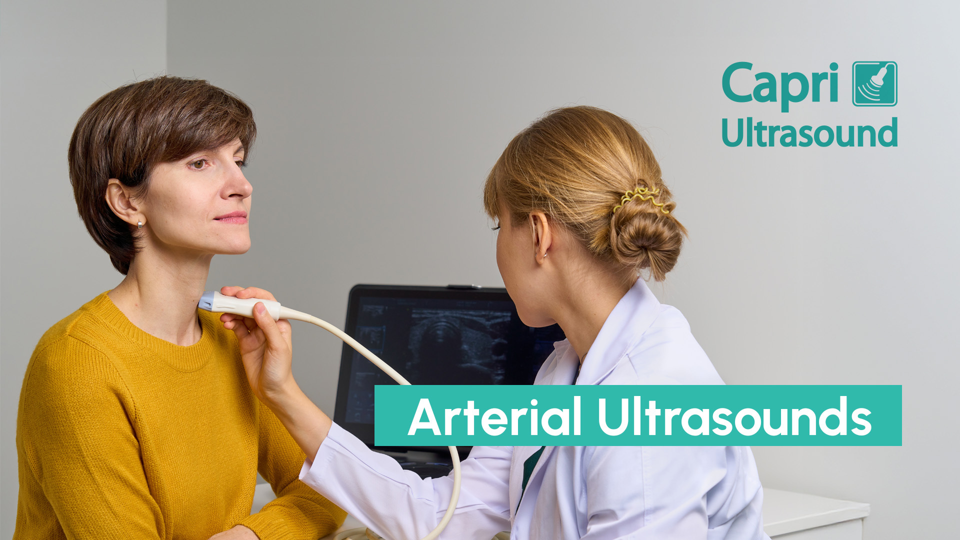
One of the most common uses for arterial ultrasound is assessing the carotid arteries which are the major vessels in your neck that supply blood to the brain. If these arteries become narrowed or blocked by plaque, it increases the risk of stroke or transient ischaemic attacks (TIAs), sometimes called “mini strokes.”
Your doctor may recommend a Doppler ultrasound of the carotid artery if you have symptoms such as:
This scan can identify issues early so that treatment can begin before a major event occurs.
Abdominal Aortic Aneurysm (AAA)
An abdominal aortic aneurysm is a dangerous enlargement of the aorta which is the body’s main artery as it runs through the abdomen. AAAs can exist silently for years and are often only detected through proactive imaging like an abdominal artery ultrasound.
If undiagnosed, an AAA may grow large enough to rupture, which can be life-threatening. Risk factors include:
Capri Ultrasound routinely includes assessment of the aorta when scanning arteries in the legs, making it an important part of preventive vascular care.
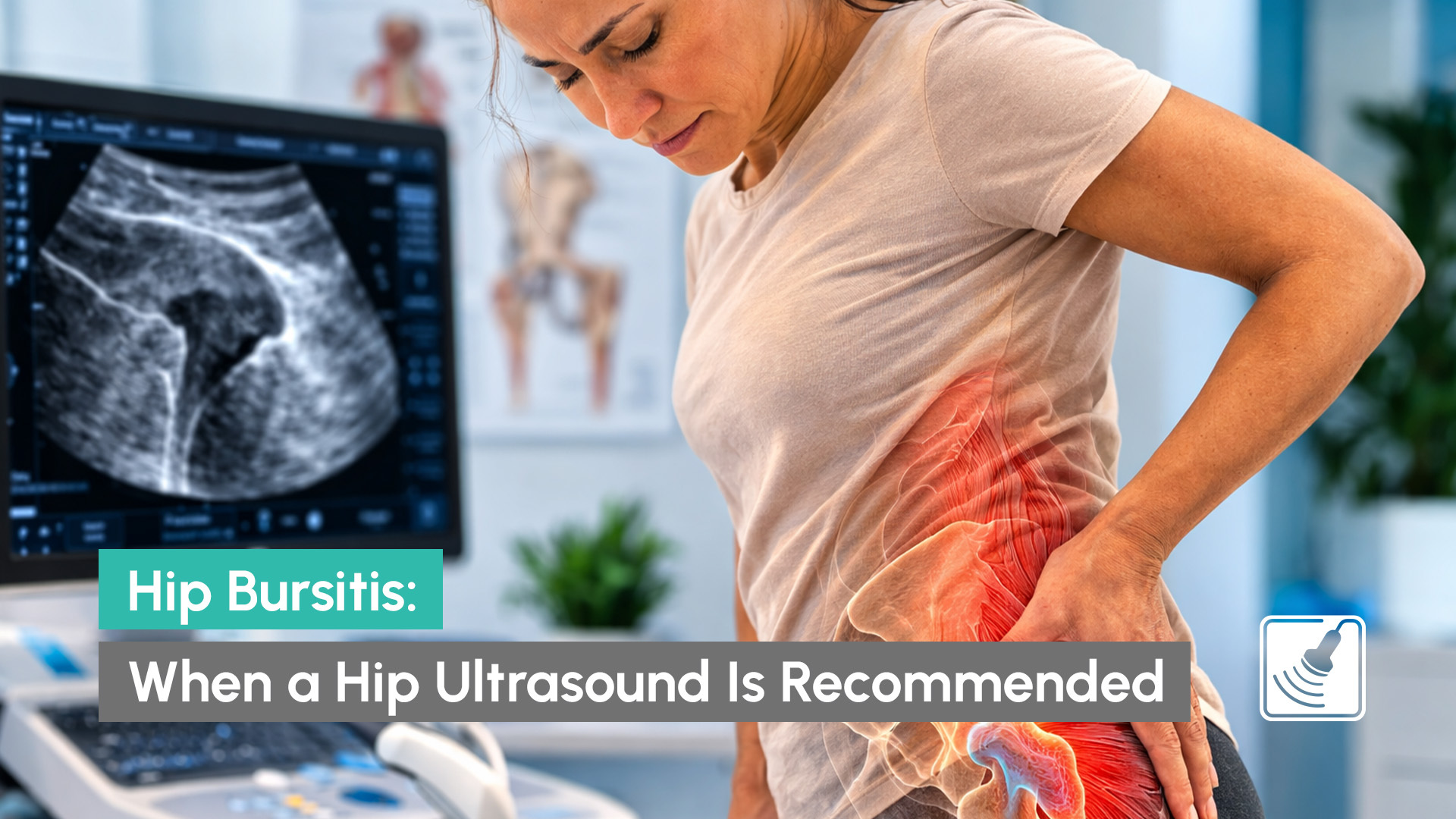
Renal Artery Stenosis & Hypertension
When the arteries that supply blood to the kidneys become narrowed, a condition known as renal artery stenosis, it can lead to persistent or resistant high blood pressure (hypertension). This is a lesser-known but important cause of secondary hypertension, especially in younger patients or those who aren’t responding to medication.
A renal artery ultrasound can help determine whether narrowed arteries are contributing to your elevated blood pressure.
Your GP or specialist may refer you for an arterial ultrasound if you have:
Early detection of arterial disease can significantly improve treatment outcomes and reduce risk of serious complications.
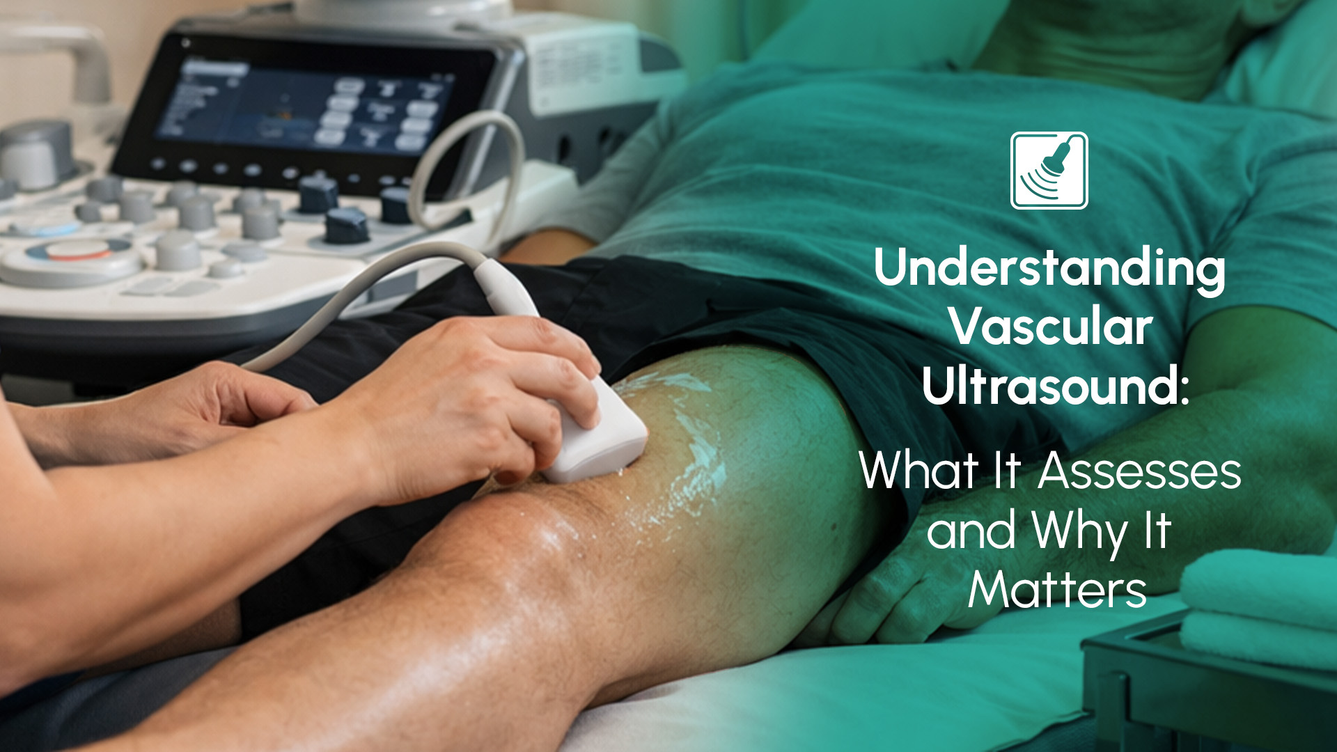
The procedure is straightforward. You’ll lie down while a trained sonographer applies a special gel to the area being examined. A small handheld device called a transducer is moved over your skin, capturing real-time images of your arteries and blood flow.
Depending on which part of the body is being scanned (neck, abdomen, or kidneys) the appointment usually takes between 10 to 20 minutes. You can eat, drink, and return to normal activities right after your scan unless advised otherwise by your doctor.
Capri Ultrasound is a trusted diagnostic imaging clinic on the Gold Coast, offering arterial ultrasound services tailored to your specific health concerns. With over 18 years of experience including working with vascular surgeons, we perform detailed vascular imaging for:
Our clinic provides expert, non-corporate care, and bulk billing is available for eligible Medicare referrals.
If you’ve been referred for an arterial ultrasound, you deserve fast, accurate, and personalised care. Capri Ultrasound offers advanced vascular imaging with bulk billing options and quick report turnaround.
📞 Contact Capri Ultrasound today to book your scan and take control of your vascular health.