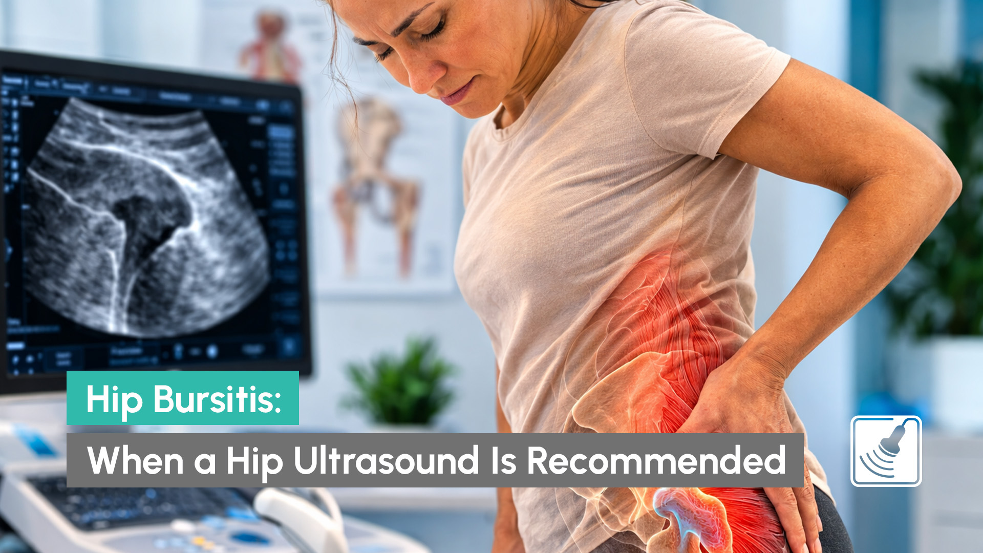
If you’re experiencing pain on the outside of your hip, especially when lying on your side or walking, your doctor may suspect hip bursitis. This condition occurs when a small fluid-filled sac on the outside of the hip becomes inflamed. In many cases, a hip ultrasound can help confirm whether hip bursitis or another soft tissue problem is causing the symptoms.
Because several conditions can cause similar pain, imaging is often used to better understand what’s happening in the hip.
Hip bursitis occurs when one of the bursae in the hip becomes irritated or inflamed. Bursae are small sacs filled with fluid that reduce friction between bones, tendons, and muscles.
The most common type affecting the hip is trochanteric bursitis, which occurs near the bony part on the outside of the hip called the greater trochanter.
When this area becomes inflamed, it can lead to symptoms such as:
Because these symptoms overlap with other hip conditions, doctors may request imaging to help confirm whether hip bursitis is the underlying cause.
The bursa located near the outer hip helps tendons glide smoothly over bone during movement. When the bursa becomes irritated, everyday activities such as walking, running, or even sleeping on your side can become uncomfortable.
Many people first notice outer hip pain that worsens after physical activity or long periods of standing. Others may experience hip pain when walking or discomfort after exercise such as running. In some cases, people report hip pain after running or other repetitive activities that place stress on the tendons surrounding the hip.
Because the outside of the hip contains several tendons and soft tissues, diagnosing the exact cause of pain can sometimes require imaging.
A hip ultrasound is commonly used to assess soft tissue structures around the hip joint.
Unlike X-rays, which show bones, ultrasound can visualise:
During the scan, a sonographer uses a small handheld probe to examine the area around the hip. In cases of hip bursitis, the inflamed bursa may appear enlarged or contain excess fluid.
A hip bursa ultrasound can also help identify other conditions that may mimic hip bursitis, such as tendon problems or muscle injuries.
Because the scan shows real-time movement of tissues, it can provide useful information about how the hip structures behave during motion.
When patients present with lateral hip pain or hip pain when lying on side, doctors often begin with a clinical examination. However, if symptoms persist or the cause is unclear, imaging may be recommended.
A hip ultrasound is particularly useful because it can:
For this reason, a hip bursitis on ultrasound assessment is often requested when symptoms suggest trochanteric bursitis or other soft tissue problems. Ultrasound is also a safe and non-invasive imaging technique that does not involve radiation.
The term trochanteric bursitis refers specifically to inflammation of the bursa located over the greater trochanter on the outside of the hip.
This condition is one of the most common causes of outer hip pain and hip bursitis symptoms.
It may develop due to:
If you’d like to learn more about bursitis in general, including causes, symptoms, and treatment options, Healthdirect Australia provides a helpful overview on Bursitis here. This resource explains the condition in more detail and when medical assessment may be recommended.
Your doctor may recommend a hip ultrasound if you are experiencing:
Because many hip conditions produce similar symptoms, imaging can help confirm whether hip bursitis or another soft tissue issue is responsible.
If your doctor has referred you for a hip ultrasound, choosing a clinic experienced in musculoskeletal imaging can help ensure accurate assessment.
Capri Ultrasound, located on the Isle of Capri on the Gold Coast, provides diagnostic
musculoskeletal scans including hip ultrasound examinations used to investigate symptoms such as hip bursitis, lateral hip pain, and hip pain when lying on side.
With a valid referral from a GP or specialist, many scans are also eligible for bulk billed ultrasound appointments.
If you’ve been experiencing persistent outer hip pain or symptoms suggesting hip bursitis, a diagnostic ultrasound at Capri Ultrasound can help determine a diagnosis. To learn more or book an appointment, contact Capri Ultrasound on Gold Coast.